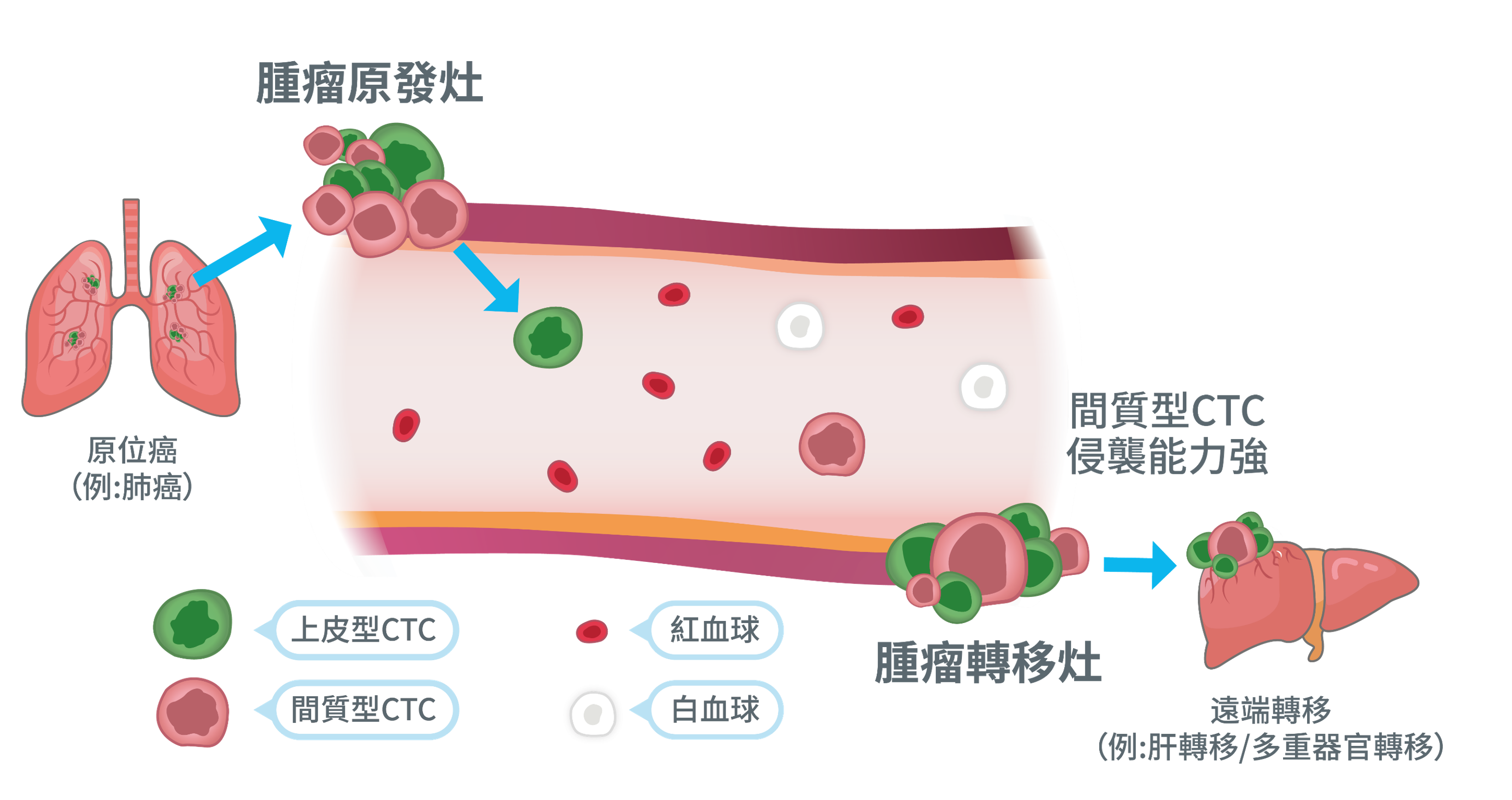
circulating tumor cells comes from cancer cells that break away from the primary tumor and enter the bloodstream, but they don't look alike. For most cancers of epithelial origin, during the process of tumor cells entering the circulation, crossing blood vessels, and forming metastases, they often appear to be epithelial, mesenchymal, or a mixture of both.
This variation is where many tests tend to miss. If the process relies only on a single epithelial marker for forward capture, it may not be fully preserved when cells lose their original epithelial characteristics and shift to a more mobile phenotype during transfer. The final results seen may not represent the actual risk profile in the sample.
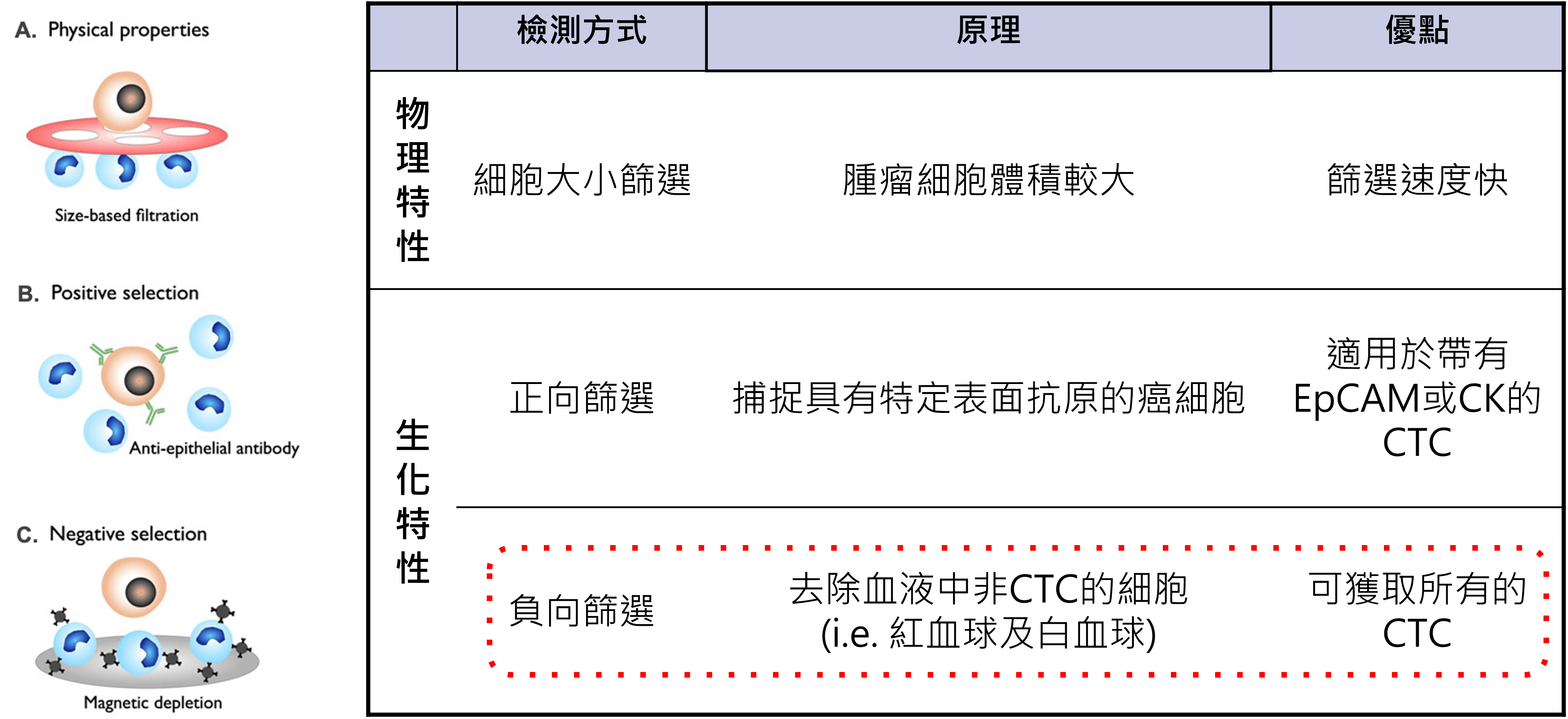
The reason why Good Future does not limit the detection to a single positive signal is because the value of CTCs lies not only in “the presence or absence” of CTCs, but in what type of cells are present in the blood. First, negative screening is used to retain more non-hematocyte targets, and then coupled with immunofluorescence staining and complete cell images, it helps to return to cell type, marker combination and signal distribution for interpretation.
For risk assessment, treatment tracking, and relapse monitoring, this information density is more important than a single number. Because what really needs to be answered is not just whether the cells exist, but what kind of tumor changes these cells may reflect, and whether they are worthy of further tracking and discussion.